Translate

Friday, 23 February 2018

Link between flu and heart attack confirmed in new research

Image result for flu and heart attackChances of a heart attack are increased six-fold during the first seven days after detection of laboratory-confirmed influenza infection, according to a new study by researchers at the Institute for Clinical Evaluative Sciences (ICES) and Public Health Ontario (PHO).
"Our findings are important because an association between influenza and acute myocardial infarction reinforces the importance of vaccination," says Dr. Jeff Kwong, a scientist at ICES and PHO and lead author of the study.
In the study published in the New England Journal of Medicine, the researchers found a significant association between acute respiratory infections, particularly influenza, and acute myocardial infarction.
The risk may be higher for older adults, patients with influenza B infections, and patients experiencing their first heart attack. The researchers also found elevated risk -- albeit not as high as for influenza -- with infection from other respiratory viruses.
"People at risk of heart disease should take precautions to prevent respiratory infections, and especially influenza, through measures including vaccinations and handwashing," says Kwong.
The researchers add that patients should not delay medical evaluation for heart symptoms particularly within the first week of an acute respiratory infection.
provided by Public Health Ontario..........

Friday, 16 February 2018

Macrophages conduct electricity, help heart to beat

Image result for macrophages conduct electricity"This work opens up a completely new view on electrophysiology; now, we have a new cell type on the map that is involved in conduction," says senior author Matthias Nahrendorf, a systems biologist at Massachusetts General Hospital, Harvard Medical School. "Macrophages are famous for sensing their environment and changing their phenotype very drastically, so you can think about a situation where there is inflammation in the heart that may alter conduction, and we now need to look at whether these cells are causally involved in conduction abnormalities."
Nahrendorf showed the results to his colleagues, David Milan and Patrick Ellinor, both electrophysiologists at Massachusetts General Hospital, who responded by opening the doors to their labs. Together, the teams found that macrophages extend their cell membranes between cardiac cells and create pores, also called gap junctions, for the electrical current to flow through. The macrophages contribute by preparing the conducting heart cells for the next burst of electricity so conducting cells are able to keep up with a fast contraction rhythm.
The group will follow up by looking at whether macrophages are involved in common conduction abnormalities. There are also potential connections between macrophages and anti-inflammatory drugs, which are widely reported to help with heart disease. If macrophages do play a role in disease, the researchers say it can open up a new line of therapeutics, as these immune cells naturally consume foreign molecules in their presence and are easy to target as a result.
Revealed by Cell Press.

Cardiac macrophages found to contribute to a currently untreatable type of heart failure


Related imageThe concept of heart failure traditionally referred to a loss of the organ's pumping capacity, which is called systolic heart failure. But in HFpEF the heart retains the ability to pump or eject blood into the circulation. What is compromised is the ability of the heart muscle to relax and allow blood to flow into the left ventricle, reducing the amount of blood available to pump into the aorta. Symptoms of HFpEF are similar to those of heart failure in general, but since factors contributing to the condition are not well understood, it has been difficult to find promising therapies.
Interactions among cells within the heart -- including macrophages -- are essential to normal cardiac function but can also contribute to problems. For example, after the heart muscle is damaged by a heart attack, macrophages induce the cells called fibroblasts to generate the connective tissues that help reinforce damaged tissue. But excessive fibroblast activation can lead to the distortion and stiffening of tissues, further reducing cardiac function.
"Not only were numbers of inflammatory cardiac macrophages increased in both the mice and in humans with HFpEF, but their characteristics and functions were also different from those in a healthy heart," says Hulsmans. "Through their participation in the remodeling of heart tissue, these macrophages increase the production of extracellular matrix, which reduces diastolic relaxation. Our findings regarding the cell-specific knockout of IL-10 are the first to support the contribution of macrophages to HFpEF."
Senior author Mathias Nahrendorf, MD, PhD , of the Center for Systems Biology, adds, "Heart muscle cells and fibroblasts have been considered the major contributors to HFpEF. Our identification of the central involvement of macrophages should give us a new focus for drug development. And since macrophages naturally take up materials for disposal, inducing them to ingest drugs carried in by nanoparticles could limit their contributions to the development of HFpEF." Nahrendorf is a professor of Radiology at Harvard Medical School.

Sunday, 11 February 2018

Red wine proves good for the heart (again).........

Antioxidants found in wine have advanced stents

Antioxidant compounds found in red wine are advancing the treatment of heart disease -- the leading cause of death for both men and women in the U.S.

Image result for red wineHeart disease occurs when plaque builds up within artery walls blocking the blood flow through tissues in the body, increasing the risk of a heart attack or stroke. About 630,000 people die each year from heart disease, according to the Centers for Disease Control and Prevention.
While there is no singular cure for heart disease, there are numerous forms of treatment including lifestyle changes and surgical procedures. In one procedure called a coronary angioplasty, a surgeon inserts and inflates a tiny balloon inside a blocked or narrow artery to widen it and allow blood to flow through to the heart thereby decreasing the risk of a heart attack or stroke. This procedure often includes inserting a permanent small mesh tube to support the blood vessel called a stent.
Commercial stents can release chemotherapy agents that are toxic and can cause the blood vessel to narrow again. LSU Department of Comparative Biomedical Sciences Professor Tammy Dugas is developing a new stent that releases red wine antioxidants slowly over time that promotes healing and prevents blood clotting and inflammation. The two antioxidant compounds are resveratrol and quercetin.
Related image"By delivering red wine antioxidants during conventional angioplasty, it may be possible to prevent excess tissue from building up and the blood vessel from narrowing again as it heals," Dr. Dugas said.
In addition to the stent, Dugas and colleagues are developing a balloon coated with the same compounds to treat blood flow blockages throughout the body called peripheral artery disease. This disease which can limit the blood flow to kidneys, the stomach, arms or legs affects about 8 to 12 million Americans. However, less than 20 percent are diagnosed by a physician. Drug-coated balloons are a relatively new product, and are being developed to help interventional cardiologists treat arteries that are difficult to target with traditional angioplasty and stent treatments.
Revealed by Louisiana State University......

Tuesday, 30 January 2018

Diagnostic test helps primary care docs rule out pathologic heart murmur in kids

Related imageAlthough heart murmur in children is usually harmless (referred to as innocent murmur), in a small number of cases it is symptomatic of cardiac disease (referred to as pathologic murmur). A new study finds that disappearance of heart murmur while standing reliably rules out pathologic heart murmurs. Using an acoustic-based, non-electronic stethoscope, researchers at two French universities noted heart sound characteristics of 194 consecutive children referred to pediatric cardiologists for heart murmur, first with patients in the supine (flat on their back) position, and then for at least one minute in the standing position.

After observational data were collected, an echocardiogram was performed to assess the presence or absence of cardiac anomalies that could explain the murmur. Eight-five percent of children (n=164) referred to a cardiologist for heart murmur did not have cardiac disease. Thirty children (15 percent) had an abnormal echocardiogram that explained the heart murmur. Of 100 children (51 percent) who had heart murmur while supine but not standing, two had an organic murmur and only one required follow up. The disappearance of heart murmur while standing, therefore, excluded a pathologic murmur with a high predictive positive value of 98 percent and a specificity of 93 percent, but with a poor sensitivity of 60 percent. In an era of highly technical medicine, physical examination should remain the first step in diagnosis, according to the authors. They conclude that the disappearance of heart murmur in children upon standing is a valuable clinical test to exclude a pathologic cardiac murmur and avoid costly referral to a cardiologist.
This was revealed by American Academy of Family Physicians....

Sunday, 31 December 2017

DEEP INSIGHT INTO THE HEART.......

Related image

By no means are only elderly people at risk from heart diseases. Physically active individuals can also be affected, for example if a seemingly harmless flu bug spreads to the heart muscle. Should this remain undetected and if, for example, a builder continues with his strenuous job or an athlete carries on training, this can lead to chronic inflammation and in the worst case even to sudden death. The latest issue of the Forschung Frankfurt journal describes how modern non-invasive examinations using state-of-the-art imaging technology can reduce such risks.

Professor Eike Nagel and his 12 coworkers at the Institute for Experimental and Translational Cardio Vascular Imaging of Goethe University Frankfurt are developing better ways to predict and diagnose heart diseases. In recent years, the researchers have taken the lead in the development of a procedure that is still very new in heart scans. Nagel explains the advantages: "With the help of magnetic resonance imaging, we can look right inside the heart muscle." Blood flow to the heart muscle is visualized and shows whether there are any constrictions of the arteries supplying the heart. Experts can also spot whether the heart muscle is scarred, inflamed or displays any other anomalies.
The comparatively fast method makes it possible to examine patients at an early stage and may prevent cardiac insufficiency or even a heart attack. "Diseases such as HIV, kidney damage, rheumatic diseases or tumours often affect the heart either directly or as a side effect of therapy," says Nagel, describing groups potentially at risk. The cardiologist is convinced: "Nowadays we can treat or even cure so many diseases, but the heart suffers too and this should be carefully monitored as it mostly remains undetected."
It is concluded  that these rapid advances in imaging over the last decades fascinating: "Nowadays we can spot the slightest changes and literally get a clear picture of the heart's condition."  

Friday, 1 December 2017

Cardiorespiratory fitness is essential to reduce risk of coronary heart disease

Coronary heart disease (CHD) is a leading cause of death for men in the U.S. Both cardiorespiratory fitness (CRF) and the blood triglyceride/high-density lipoprotein ratio (TG:HDL ratio) are strong predictors of death from CHD. In the current issue of Mayo Clinic Proceedings, two new studies highlight the importance of CRF on subsequent CVD and mortality risk. These articles contribute substantive evidence on the importance of achieving moderate to high levels of CRF in both adults and children.
In an investigation led by Stephen W. Farrell, PhD, of The Cooper Institute, Dallas, TX, researchers found strong evidence that moderate-to-high level of fitness counteracted some of the negative effects of a high TG:HDL ratio.
Related image"While it is still extremely important to measure traditional risk factors such as resting blood pressure, blood cholesterol, triglyceride, and glucose levels, having a measure or estimate of the patient's cardiorespiratory fitness level gives us additional information regarding cardiovascular disease risk," explained Dr. Farrell. "The results of this study support this recommendation. Regardless of whether the blood TG:HDL ratio was low or high, having at least a moderate level of fitness provided some protection from CHD death when compared to having a low level of fitness."
A total of 40,269 men received a comprehensive physical examination between January 1, 1978 and December 31, 2010. The exam included a maximal treadmill exercise test to measure cardiorespiratory fitness level, and also included measurement of the blood TG:HDL ratio. This ratio is easily calculated by taking the fasting blood triglyceride level and dividing it by the blood HDL cholesterol level. A lower ratio is an indicator that insulin is working well, while a higher ratio indicates resistance to insulin. Higher ratios also indicate an increased risk of future prediabetes, type 2 diabetes, and cardiovascular disease.
The participants, categorized into low, moderate, and high CRF groups, were followed for an average period of 16.6 years, during which time 556 deaths due to coronary heart disease (CHD) occurred. Moderate to high levels of fitness provided significant protection from CHD death. Lower values for the TG:HDL ratio also provided significant protection. The lowest risk of CHD death was seen among high fit men in the lowest category of TG:HDL ratio, while the highest risk of CHD death was seen among low fit men in the highest category of TG:HDL ratio. Within each of the four categories of TG:HDL ratio, having a moderate to high level of fitness provided significant protection against CHD death when compared to having a low level of fitness. Therefore, knowing the patient's fitness level as well as their TG:HDL ratio provides much more information about CHD risk status than just knowing one or the other. When used in combination with other risk factors, such as the patient's blood pressure, bloodwork, family history, etc., measuring or estimating the patient's level of CRF can result in a much more accurate determination of their cardiovascular disease risk status.

Thursday, 23 November 2017

Uninsured heart attack, stroke patients face ‘catastrophic’ costs

"Many patients with large medical bills may have had to declare bankruptcy, sell their home, and be hounded by medical bill collectors as they struggled to juggle rent or mortgage payments while paying for utilities and food," said cardiologist Dr. Rohan Khera, an Internal Medicine fellow at UT Southwestern Medical Center and co-first author of the paper, "Medical expenses are the leading cause of bankruptcy in the United States."

Image result for heart attackFor uninsured patients hospitalized with acute myocardial infarction (heart attack), the median cost for their stay totaled $53,384 in 2012, according to the research. Uninsured stroke patients had a median bill of $31,218.
About 60 percent of the uninsured heart attack patients had bills higher than their annual income, after allowing for the purchase of food, according to the paper.
The research, published in the journal Circulation, looked at thousands of patients hospitalized for acute myocardial infarction (heart attack) or stroke between 2008 and 2012, the year before Americans began signing up for subsidized insurance on exchanges created under the federal Affordable Care Act (ACA).
The patients studied ranged from age 18 to 64 (federal Medicare insurance is available to those 65 and older). More than 39,000 hospitalizations for heart attack and more than 29,000 hospitalizations for stroke were analyzed.
Information for the paper came from the National Inpatient Sample, a large database developed by the federal Agency for Healthcare Research and Quality.
Collaborators included cardiologist Dr. James de Lemos, Professor of Internal Medicine and holder of the Sweetheart Ball‐Kern Wildenthal, M.D., Ph.D. Distinguished Chair in Cardiology and Medical Director for the Dallas Heart Study, and researchers were from Baptist Health South Florida, Johns Hopkins Bloomberg School of Public Health, Florida International University, Michael E. DeBakey VA Medical Center, Brigham and Women's Hospital, and Yale New Haven Hospital.

Thursday, 16 November 2017

SURFACE ANATOMY OF HEART

Orientation and Surfaces

The heart has been described by many texts as “a pyramid which has fallen over”. The apexof this pyramid pointing in an anterior-inferior direction.
In its typical anatomical orientation, the heart has 5 surfaces, formed by different internal divisions of the heart:
  • Anterior (or sternocostal) – Right ventricle.
  • Posterior (or base) – Left atrium.
  • Inferior (or diaphragmatic) – Left and right ventricles.
  • Right pulmonary – Right atrium.
  • Left pulmonary – Left ventricle.

Borders

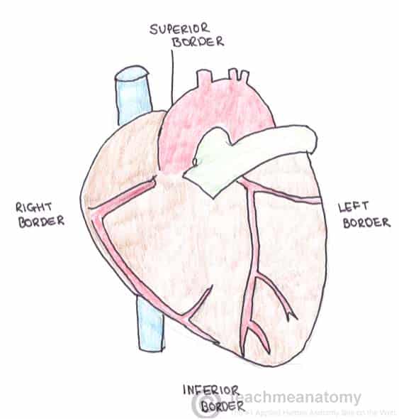
Separating the surfaces of the heart are its borders. There are four main borders of the heart:
  • Right border – Right atrium
  • Inferior border – Left ventricle and right ventricle
  • Left border – Left ventricle (and some of the left atrium)
  • Superior border – Right and left atrium and the great vessels
Fig 1.1 - Borders of the heart.

Fig 1.0 – Borders of the heart.

Sulci of the Heart

The heart is a hollow structure. On the interior, it is divided into four chambers. These divisions create grooves on the surface of the heart – these are known as sulci.
The coronary sulcus (or atrioventricular groove) runs transversely around the heart – it represents the wall dividing the atria from the ventricles. The sinus contains important vasculature, such as the right coronary artery.
The anterior and posterior interventricular sulci can be found running vertically on their respective sides of the heart. They represent the wall separating the ventricles.

Pericardial Sinuses

The pericardial sinuses are not the same as ‘anatomical sinuses’ (such as the paranasal sinuses). They are passageways formed the unique way in which the pericardium folds around the great vessels.
  • The oblique pericardial sinus is a blind ending passageway (‘cul de sac’) located on the posterior surface of the heart.
  • The transverse pericardial sinus is found superiorly on the heart. It can be used in coronary artery bypass grafting – see below.

Heart Conduction..........

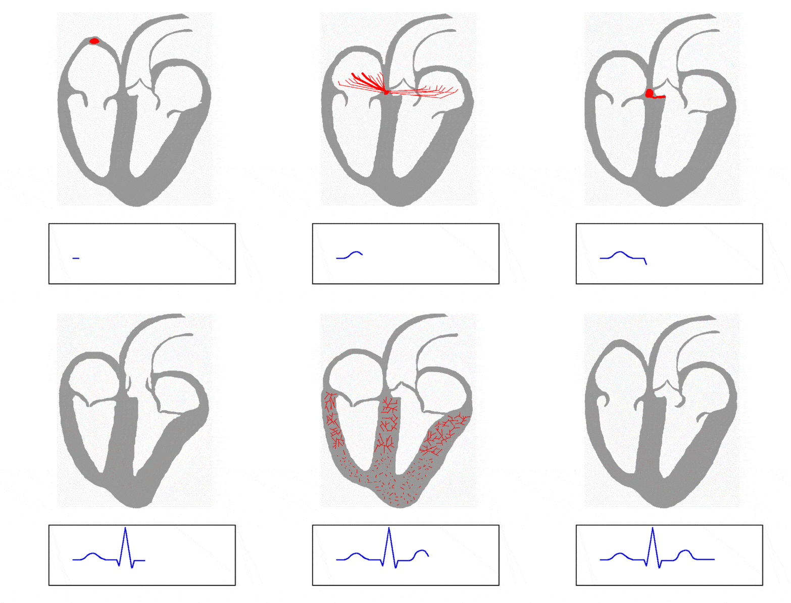
The sequence of electrical events during one full contraction of the heart muscle:
    Fig 1.0 - Animation of the spread of conduction through the heart
  • An excitation signal (an action potential) is created by the sinoatrial (SA) node.
  • The wave of excitation spreads across the atria,causing them to contract.
  • Upon reaching the atrioventricular (AV) node, the signal is delayed.
  • It is then conducted into the bundle of His, down the interventricular septum.
  • The bundle of His and the Purkinje fibres spread the wave impulses along the ventricles, causing them to contract.

Thursday, 26 October 2017

BLOOD PURIFIERS......

1. Water

The standard recommendation for drinking water is 8 glasses of water But drink as much as you need.
Image result for water glassThis should come as no surprise to you. Since urine is the means through which kidneys flush out toxins from the blood, having sufficient water is your best bet to keep the blood clean. As with a deep cleanse of your home, a proper purification regimen for your blood will require you to incorporate plenty of water. Staying hydrated can help your kidneys flush out the impurities in the body, aided by the fiber and minerals from all the other blood purifying foods.
Ayurveda outlines an ideal way to have water for detox benefits. Keep some warmed water in a copper vessel overnight and drink it next day morning when you wake up. The copper cools your liver and the water helps with waste elimination and purification of the blood.

2. Garlic

Garlic is antibacterial and can cleanse your system of viruses and parasites that could harm it. This pungent food removes excess fat from the blood. In the bargain, it lowers the levels of bad LDL cholesterol and total serum cholesterol in your body.
Lead is a common toxin lurking in paint and other household objects that pollutes the blood. Garlic can bring down lead levels by 19%.
Image result for garlicExposure to toxic metals can pollute your blood and body, causing all manner of problems – from cramps and appetite loss to brain damage. The antioxidants in garlic not only detoxify your body as a whole but can also fight mild-to-moderate lead poisoning. You are often exposed to this metal through lead paints, contaminated dust, etc. Garlic can reduce metal levels by as much as 19% and ease toxicity symptoms like headaches. Research has shown that the sulfur compounds in garlic have a therapeutic effect not unlike that of d-penicillamine (used for treating lead poisoning). Garlic could, in fact, be a safer option to cleanse your body of the heavy metal.

3. Jaggery

Image result for jaggery
Though sweet, jaggery helps detox the blood.
A popular ingredient from India, this golden-brown unrefined sugar is also known to be a good blood purifier. Its fiber content helps it cleanse the digestive system, prevent constipation, and expel waste from the body. Because of the high iron content, it can help sort out an iron deficiency and restore hemoglobin levels. If you’ve just had a baby, jaggery can help expel clotted blood from the body, acting as a natural cleanse/flush for your blood.

4. Turmeric

Turmeric on its own is a wonderful natural healer that can fight inflammation. It can help treat liver trouble and even keep it functioning optimally. And this is important because the liver and kidneys are the two primary centers for cleansing the blood of impurities and purging them from the body.
Infuse black pepper, cardamom, cinnamon, cloves, and ginger in a glass of turmeric milk for blood-purifying benefits.
Image result for turmericWhen this golden spice is combined with milk, its purification powers go up a notch. Turmeric milk figures prominently in liver cleanse diets in Ayurveda. One such diet recommends turmeric milk that has been infused with spices like black pepper, cardamom, cinnamon, clove, and ginger for 72 hours. This drink is supposed to help your body generate fresh red blood cells besides plying your system with nutrients and acting as a good health tonic.

5. Fresh Fruit Like Apples, Pears, And Guavas

Don’t peel fruits like apples and guava. The peel is a richer source of antioxidants.
Image result for appleThe pectin fiber in fruit like apples, guavas, plums, and pears is useful for detoxifying the blood. Besides binding with the excess fats in your blood as well as liver, pectin also combines with heavy metals and other harmful chemicals or waste in your blood stream. The fiber helps with the fat removal, while the lycopene and glutathione in the fruits are useful for eliminating waste and chemicals. Lycopene is found in red fruits like watermelon and pink guava while apples are a good source of glutathione.

6. Lemon

Warm water with lemon juice is recommended in both Ayurveda and naturopathy as a good start-of-day drink to cleanse the blood. As per Ayurveda, this helps release the “ama” or toxins in your digestive tract, cleansing your system.
Image result for lemon
A warm glass of lemon water in the morning can increase glutathione in your body, which helps the liver detox the blood better.
Research also backs this up. The warm water aids in the breakdown of fat and also eases the load on your kidneys and the vitamins and minerals in the lemon help the body detox. The vitamin C, in particular, is needed by your body to create glutathione. Your liver then uses this compound to purge your blood of harmful chemicals.

7. Dark Green Leafy Vegetables

Image result for spinach
Not only do leafy greens like spinach supply your blood with iron to help maintain the hemoglobin levels, they also check free radical damage to the blood cells.
Not everyone’s a fan of dark green leafy vegetables. But they can be very useful thanks to their abundant nutrient content (vitamins A and C, as well as B vitamins, folic acid, and iron). The antioxidants in these vegetables help remove harmful free radicals, responsible for cell damage to red blood cells. By limiting free radical damage to blood cells, these vegetables help keep your blood replenished with new cells.  Choose from kale, spinach, Romaine lettuce, or even mustard greens and Swiss Chard.
8. Cabbage
Cook the cabbage well if you suffer from hypothyroidism. Cooking removes the goiter-causing substances.
Image result for cabbageCabbage is considered in Ayurveda as a blood purifier. This leafy cruciferous vegetable has antioxidants like vitamins A and C that are good for your liver. The fiber in cabbage helps cleanse the digestive tract. It can neutralize chemical compounds like those in cigarette smoke and cleanse your liver, too. The glucosinolates in cabbage (which give cruciferous vegetables their distinctive taste) break down to form isothiocyanates. These help expel carcinogens in the body.

9. Oats, Wheat Bran, And Nuts

Image result for oatsThese fiber-rich whole grains and nuts are a must-have for their ability to rid your system of excess fat, chemicals, and waste. The high fiber content in foods like oats, whole grains, wheat bran, flaxseeds, and nuts lowers blood cholesterol and glucose levels, cleanses your intestinal tract, eases constipation, and helps your body eliminate waste better.

10. Watercress

Image result for watercressThis may not be your usual mealtime staple, but watercress is a delicious alternative to regular greens. It helps increase levels of detoxification enzymes in the body. It also eliminates carcinogens in the body in much the same way as cabbage does – through the action of the glucosinolates in it. In one study on the effects of watercress intake on smokers, researchers found that just 170 g a day resulted in higher levels of carcinogens being expelled in the urine, leaving the blood and body more purified.
Besides incorporating these blood-purifying foods in your diet, remember to avoid foods that increase cholesterol and triglycerides and foods that increase glucose in the blood.

World Heart Day

This year on World Heart Day, as part of our mission to ensure heart health equity for all, we want to create a global community of He...