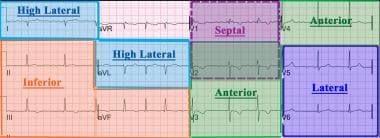
The ECG has its roots in the seminal work of Galvani, who famously noted the contraction of frog legs when stimulated with electricity. Although he was incorrect in attributing this phenomenon to "animal electricity," his findings ushered in a period of research in the interaction of electricity and biological systems.
Matteucci, and later Kolliker and Muller, demonstrated that the electrical activity of a beating heart caused synchronous contractions in muscle preparations that were connected to the heart by nerve tissue. Gabriel Lippman developed the capillary electrometer, making it possible to measure the minute currents in biological systems. With the Lippman electrometer, Agustus Waller performed the first ECG of a human heart at St Mary’s Hospital in London in May 1887. Willem Einthoven improved the ECG using the Lippman electrometer and then introduced the string galvanometer, which represented a great leap forward in electrocardiography. With Einthoven’s improvements, the now familiar P, Q, R, S, and T waves were apparent.
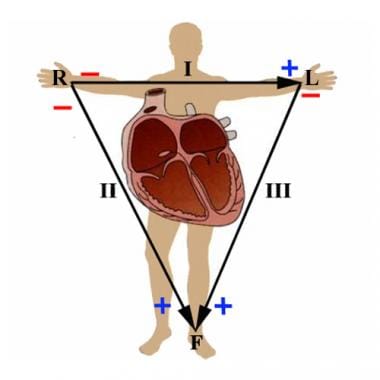
 Not only did Einthoven reveal the ECG waveforms as we know them today, but his convention for placement of the ECG leads is still in use, with leads on the right and left arms as well as the left leg, using the right leg as an electrical ground. With these leads, he demonstrated the well-known "Einthoven’s triangle".
No comments:
Post a Comment